Meet the Team

  • Mentors: Dr. Prashant Jindal, Dr. Mamta Juneja, Dr. Rakesh Tuli, Sarthak Thakur, Anuj Wani, Archit Uniyal
  • Team interns:
  • Ojaswani 
  • Amit
  • Durgesh

Introduction: Computer Aided Diagnosis System for Glaucoma using Retinal Fundus Imaging


Applications:

The propped CAD system can be used for large scale screening of Glaucoma at medical institutes and can save time by not using traditional diagnosis approaches.

Achievements

  • Juneja M, Singh S, Agarwal N, Bali S, Gupta S, Thakur N, Jindal P. Automated detection of Glaucoma using deep learning convolution network (G-net). Multimedia Tools and Applications. 2019:1-23. (Science citation index, Impact factor: 2.1)
  • DC-Gnet for detection of glaucoma in retinal fundus imaging (Communicated).
  • GC-NET for classification of glaucoma in retinal fundus image (Communicated)

Motive:

To design and develop a Computer Aided System (CAD) system based on convolutions which can identify patients with diagnosing Glaucoma at an early stage in order to ease the work of doctors.

Features:

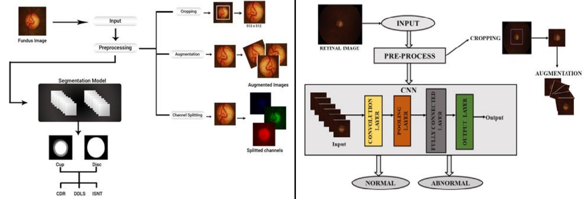
The Classification technique developed uses Convolution Neural Networks at it’s core along with Image Processing techniques to classify patients as Glaucomatous or Normal.

The CNN model takes Fundus images as an input.

The output generated is whether the patient suffers from Glaucoma or not and if yes, then up to what extent.

Uses Data Augmentation techniques in order to increase the dataset for training the CNN.

This CNN model does not require any human intervention and can function on it’s own with minimalistic human efforts.


Information

  • Name of the Department: UIET, Panjab University
  • Name of Project: Design Innovation Centre(DIC)
  • Name of Group : Medical Devices and Restorative Technologies
  • Partners: PGIMER , GMCH 32, Chandigarh
  •  

© Design Innovation All Rights Reserved

wpChatIcon